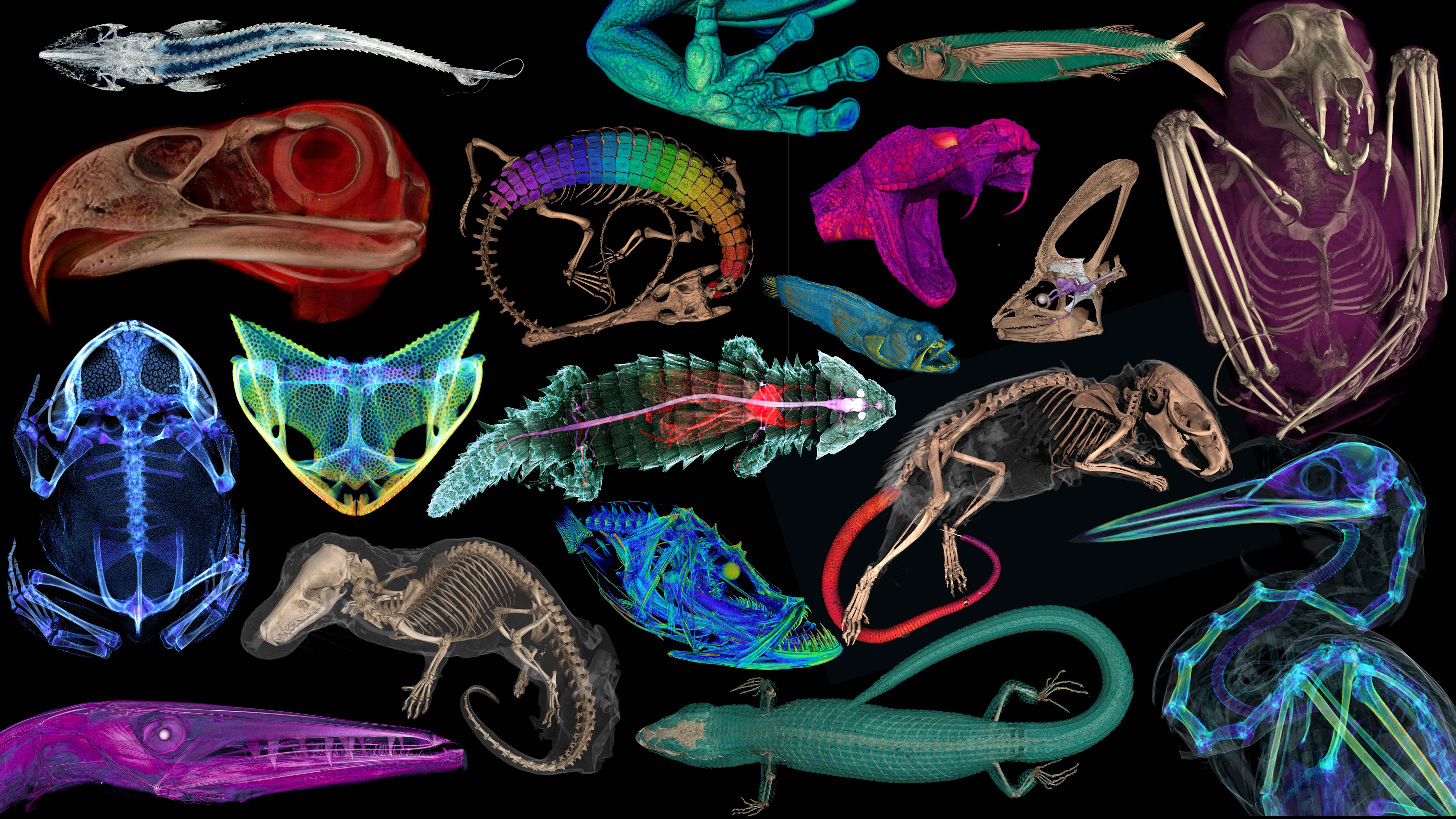
 Specimens once restricted solely to the scientists who study them are now available as 3D models to everyone. (Florida Museum image by oVert)
 CHICAGO — Natural history museums are celebrating the launch of a major digitization project that offers the public and scientists access to CT scans — detailed internal images — of some 13,000 species of animals. DePaul University mammologist Noé de la Sancha contributed to
openVertebrate (oVert), a five-year collaboration among 18 institutions to create 3D reconstructions of vertebrate specimens, now freely available online.
De la Sancha studies small mammals and is a co-author of a summary of the project,
published in the journal BioScience. Co-authors review the specimens that have been scanned to date and offer a glimpse of how the data might be used to ask new questions and spur the development of innovative technology.
“Many museums are housing individual mice that are extremely important and one of a kind. Sometimes it’s a mouse someone found in the early 1900s,” de la Sancha said. “This project will help to democratize access to specimens from around the world.” Scanning the rodent tree of life
As part of his contribution to the project, de la Sancha and Cody Thompson at the University of Michigan earned a Partner to an Existing Network grant from the National Science Foundation. This initiative,
Digitization PEN: Functional Quantitative Characters for Ecology and Evolution (FuncQEE), tackles a subsection of the vertebrate tree of life that includes rodents.
While CT scans of animals have existed for years, few wildlife species were represented or available for study. Dissected animals fall apart, while CT scans illuminate body systems intact and reveal details scientists could not see before. For de la Sancha, this means measuring how species have changed over time and space. De la Sancha’s team set out to identify functional traits for rodents throughout the
rodent tree of life within the oVert project.
“Rodents are the most divers group of mammals alive,” says de la Sancha, an assistant professor of environmental science and studies. “We wanted to come up with functional traits that we could quantify, and CT scanning allows us to do this in a way we never could before.”
The initiative creates huge amounts of data, which provides both opportunities and challenges. It can be costly to store and process large files, noted de la Sancha. Continued developments in machine learning and supercomputing will be essential as scientists seek to use artificial intelligence to unlock the images’ full potential.
“The data will help us approach questions about ecology and evolution, including why physical traits allow certain species to survive in different types of environments, which can be used to better understand patterns of biodiversity especially in quickly changing habitats. For example, the Atlantic Forest of South America which has been experiencing severe deforestation in the past decades,” said de la Sancha.
Team project spans years
Between 2017 and 2023, oVert project members took CT scans of more than 13,000 specimens, with representative species across the vertebrate tree of life. This includes more than half the genera of all amphibians, reptiles, fishes and mammals. CT scanners use high-energy X-rays to peer past an organism’s exterior and view the dense bone structure beneath. Thus, skeletons make up the majority of oVert reconstructions. A small number of specimens were also stained with a temporary contrast-enhancing solution that allowed researchers to visualize soft tissues, such as skin, muscle and other organs.
oVert was funded with an initial sum of $2.5 million from the National Science Foundation, along with eight additional partnering grants totaling $1.1 million that were used to expand the project’s scope. The goal was to initially scan only specimens preserved in ethyl alcohol, which represent the bulk of fish, reptile and amphibian collections.
Soon, the project grew to include a scan of a humpback whale, spinosaurus and more. Edward Stanley is co-principal investigator of the oVert project and associate scientist at the Florida Museum of Natural History. In 2023, he was conducting routine CT scans of
spiny mice and was surprised to find their tails were covered with an internal coat of bony plates, called osteoderms. Before this discovery, armadillos were considered to be the only living mammals with these structures.
“All kinds of things jump out at you when you’re scanning,” Stanley said. “I study osteoderms, and through kismet or fate, I happened to be the one scanning those particular specimens on that particular day and noticed something strange about their tails on the X-ray. That happens all the time. We’ve found all sorts of strange, unexpected things.”
For his part, de la Sancha has shuttled preserved specimens between The Field Museum in Chicago to CT scanners at the University of Michigan. De la Sancha’s work continues as the team wrap ups their FuncQEE project this year. He plans to incorporate the project into his teaching and research, training students at DePaul to use the project’s data.
Learn more about the
oVert project online.
Portions of this press release were written by Jerald Pinson of the Florida Museum of Natural History.
###
Source:
Noé de la Sancha
ndelasa1@depaul.edu
Media Contact:
Kristin Claes Mathews
kristin.mathews@depaul.edu
312-362-7735